When you are diagnosed with wet AMD and at future visits to your eye doctor, you may receive an OCT (optical coherence tomography) scan1. OCT scans are a non-invasive way to take a picture of the back of your eye. These pictures help detect changes in your condition, including the presence or absence of fluid1.
Two main types of fluid your doctor may find on the scan are intra-retinal fluid (IRF) and sub-retinal fluid (SRF)2. Build-up of these fluids in your eye may be responsible for blurry, wavy or distorted central vision2,3.
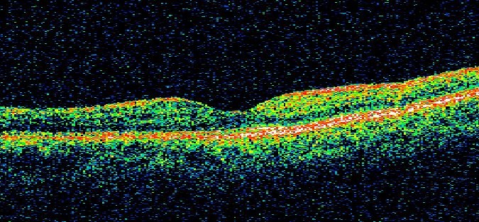
Here is an example of an OCT scan of a patient with wet AMD:

Your eye doctor will take OCT scans regularly during your visits and check them for fluid1. When treatment for wet AMD is effective, little to no fluid will be present in the back of the eye4.
The following OCT image has no fluid present in the back of the eye:
-
Kang SW, et al. The correlation between fluorescein angiographic and optical coherence tomographic features in clinically significant diabetic macular edema. Am J Ophthalmol 2004;137(2):313-322.
-
Arnold J et al. The role of sub-retinal fluid in determining treatment outcomes in patients with neovascular age-related macular degeneration–a phase IV randomised clinical trial with ranibizumab: the FLUID study. BMC Ophthalmol. 2016;143(4):679-680.
-
National Eye Institute. Facts About Age-Related Macular Degeneration. Available at https://nei.nih.gov/health/maculardegen/armd_facts (link is external). Accessed August 2019.
-
García-Layana A, et al. Early and intermediate age-related macular degeneration: update and clinical review. Clin Interv Aging. 2017; 12:1579–1587.