Test at home today using the Amsler grid
Because wet macular degeneration is a chronic, degenerative condition, you, your caregiver(s) and eye doctor will all have a role in monitoring its progression when you are on an anti-VEGF treatment.
How you can monitor the progression of wet macular degeneration1,2,3
Your eye doctor may ask you to keep track of your vision using a black and white grid called an “Amsler” grid.
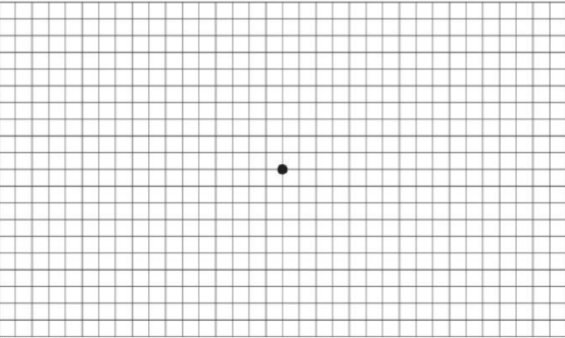

Caption: Amsler grid as seen without wet macular degeneration (left); Amsler grid as seen with wet macular degeneration (right)
Here is how to test your vision with an Amsler grid:
- CLICK HERE to download a grid and print it on your home printer.
- Go to the room in your home where you do most of your reading, and use the light you normally use to read
- Wear eyeglasses you normally wear for reading
- Place or hold the Amsler grid approximately 30 to 38 centimeters, or 12 to 15 inches, from your eyes
- Test each eye individually: Cover one eye while testing the other eye
- Keep your eye focused on the dot in the center of the grid and answer these questions:
- Do any of the lines in the grid appear wavy, blurred or distorted?
- Do all the boxes in the grid look square and the same size?
- Are there any “holes” (missing areas) or dark areas in the grid?
- Can you see all corners and sides of the grid (while keeping your eye on the central dot)?
- Switch to the other eye and repeat.
It is important to report any irregularities to your eye doctor immediately. Mark areas of the Amsler grid that you’re not seeing properly and bring the grid with you when you visit your eye doctor.
Check your eyes with the Amsler grid as frequently as your doctor recommends, or whenever you notice a change in your eyesight.
How your eye care professional monitors the progression of wet macular degeneration4
Your eye doctor will likely monitor the progression of your wet macular degeneration using eye exams and by taking special pictures of the back of your eye using an OCT (optical coherence tomography) scan. A common, noninvasive technique that produces images of the back of the eye, the OCT will help your eye doctor monitor changes in your condition, including the presence or absence of fluid.
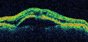
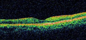
Caption: OCT image of eye with wet macular degeneration (left); OCT image of healthy eye (right)
References
- Schwartz R, Loewenstein A. Early detection of age related macular degeneration: current status. Int J Retina Vitreous. 2015;1:20.
- American Academy of Ophthalmology. Have AMD? Save Your Sight with an Amsler Grid. Available at: https://www.aao.org/eye-health/tips-prevention/facts-about-amsler-grid-daily-vision-test. Accessed July 2019.
- American Macular Degeneration Foundation. Amsler Chart to Test Your Sight. Available at: https://www.macular.org/amsler-chart. Accessed July 2019.
- Kang SW, et al. The correlation between fluorescein angiographic and optical coherence tomographic features in clinically significant diabetic macular edema. Am J Ophthalmol 2004;137(2):313-322.