The most important eye exams – with a special focus on retinal conditions
As we grow older, the risk of being diagnosed with a vision loss condition increases. Regular examinations are vital to ensure detection and treatment of such conditions, including wet age-related macular degeneration.1,2 With so many tests available, it can be overwhelming to keep track of the most important ones. Below is a brief list of some of the most common and critical eye examinations for vision loss conditions like wet AMD. If there are features of AMD present during your routine eye check, you will be referred to an ophthalmologist for further investigations and treatment.
First things first: what happens during a visit to your ophthalmologist before testing?
When you first arrive for your appointment you will typically be pre-examined by a medical technician or eyecare nurse before speaking with your ophthalmologist. In the case of wet age-related macular degeneration (wet AMD), this pre-examination may include an Amsler grid test which can help detect and monitor wet AMD.3
After meeting with a medical technician or eyecare nurse, you will speak with your ophthalmologist about specific vision challenges or other conditions, like diabetes or high blood pressure, which may affect your vision. During the discussion, the ophthalmologist determines further eye exams to be performed. The methods your ophthalmologist uses for your exam depend on your individual patient history.4
Standard eye exams: vision tests
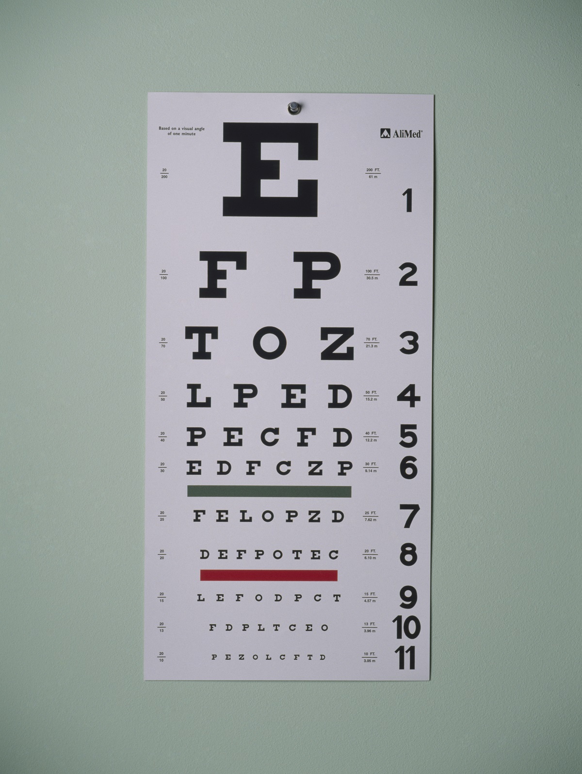
Exams with your ophthalmologist typically begin with classic vision tests. The ophthalmologist will use a vision chart showing letters or numbers of decreasing sizes (called an ETDRS vision chart). You will be asked to read the lines of letters or numbers, while covering one eye and sit facing the chart at various distances. They may also show you a chart with C-shaped rings (Landolt rings). The C-shaped rings will be in different positions and sizes, and during the test you will be asked which side of the ring is open.4
Examinations of the retina
If damage to your retina is suspected, the ophthalmologist will initiate further testing. This can include the following methods:
- Fundoscopy: Using a special light and a magnifying lens, the physician examines the back of the eye and the retina. Here, drusen (deposits below the retina), which are commonly present with wet AMD may be detected.5
- Fluorescein angiography: Fluorescein dye is injected into the arm and photographs of the eye are taken to show the fine structure of the retina, to detect newly formed blood vessels indicative of wet macular degeneration or other retinal conditions.6
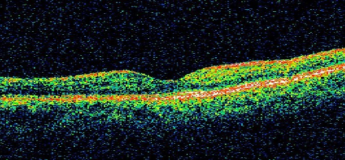
- Click here to learn about Optical coherence tomography (OCT). During this exam, laser scans of the retina, including the macula, are taken to see the layers of the retina. For wet macular degeneration, these results are used to detect fluid in this area.6
In case of a retinal condition diagnosis, your ophthalmologist will perform regular vision tests and OCT exams to inform treatment decisions. Fluorescein angiography is often only performed at diagnosis.6
After visiting your ophthalmologist: What you can do at home?
You should periodically check your vision in between appointments to monitor for any differences to report to your doctor. Amsler grids are simple to use at home so that any changes can be addressed quickly.7 Ask your eye doctor if they have any support materials for you.
It is important to take advantage of every single ophthalmologist appointment to optimise your treatment and help maintain your independence. For tips on preparing for appointments, click here.
-
Bressler NM. Early detection and treatment of neovascular age-related macular degeneration. The Journal of the American Board of Family Practice, 2002;15(2):142-152
-
Schmidt-Erfurth U, et al. Guidelines for the management of neovascular age-related macular degeneration by the European Society of Retina Specialists (EURETINA). Br J Ophthalmol 2014; 98:1144–1167. doi:10.1136/bjophthalmol-2014-305702
-
Schwartz R, Loewenstein A. Early detection of age-related macular degeneration: current status. Int J Retin Vitr (2015) 1:20. 2015. DOI 10.1186/s40942-015-0022-7
-
Turbert D. Eye Exam and Vision Testing Basics. American Academy of Ophthalmology, 17 Mar. 2020, aao.org/eye-health/tips-prevention/eye-exams-101.
-
Kang SW, et al. The correlation between fluorescein angiographic and optical coherence tomographic features in clinically significant diabetic macular edema. Am J Ophthalmol 2004;137(2):313-322.
-
American Academy of Ophthalmology. Have AMD? Save Your Sight with an Amsler Grid. Available at: https://www.aao.org/eye-health/tips-prevention/facts-about-amsler-grid-daily-vision-test. Accessed July 2019.